










































































Your cart
There are no more items in your cart


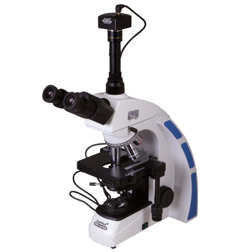
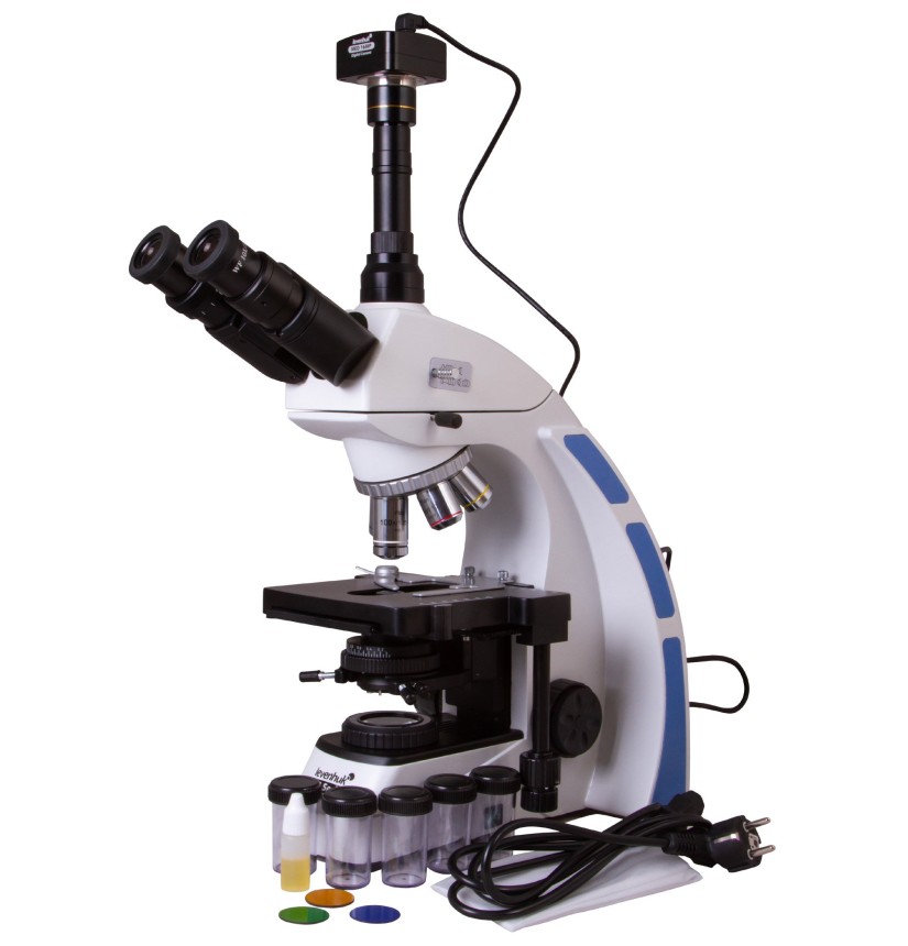
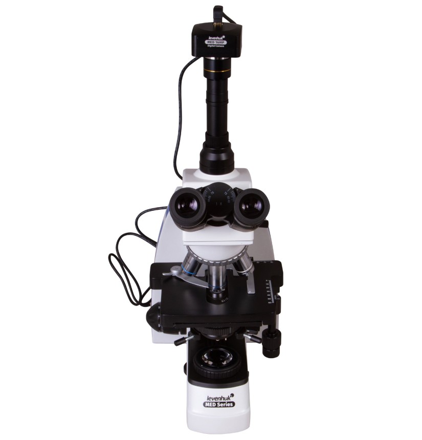
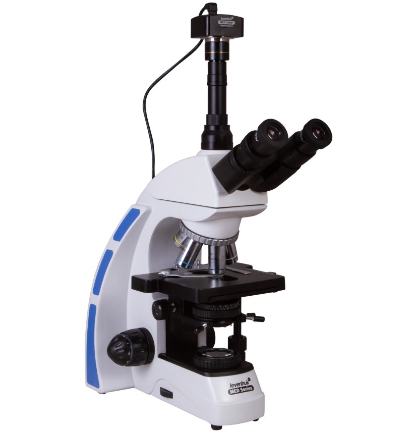
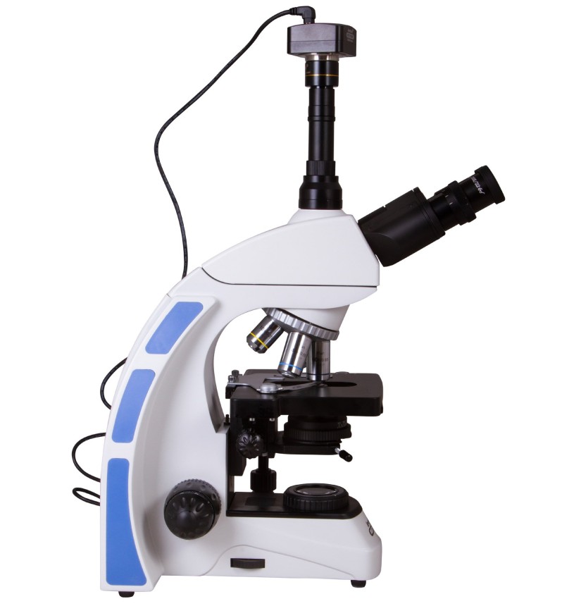
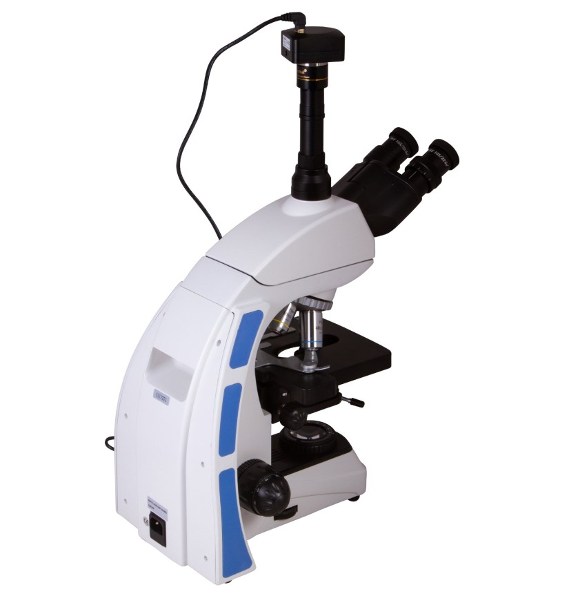
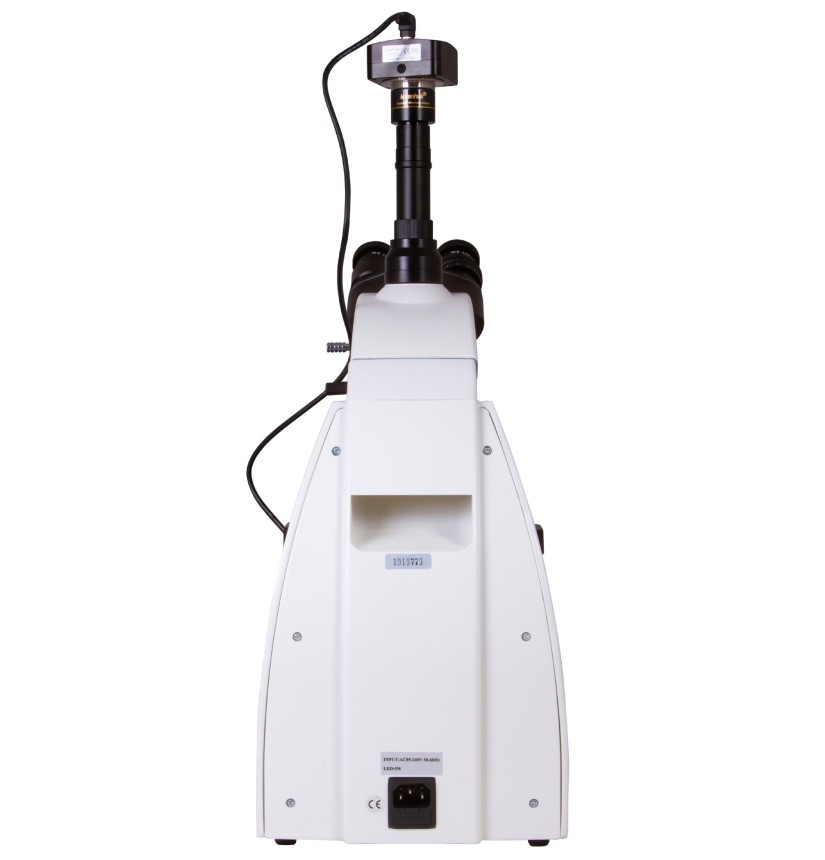
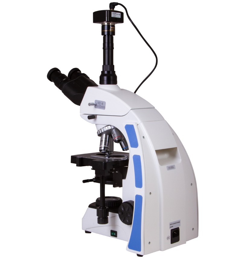
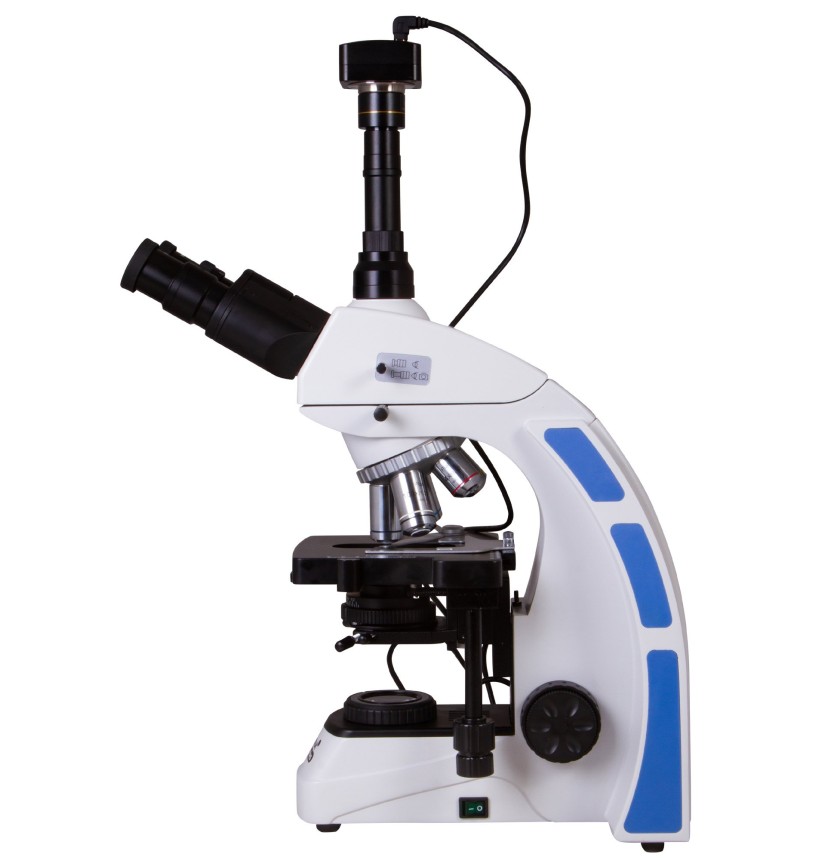
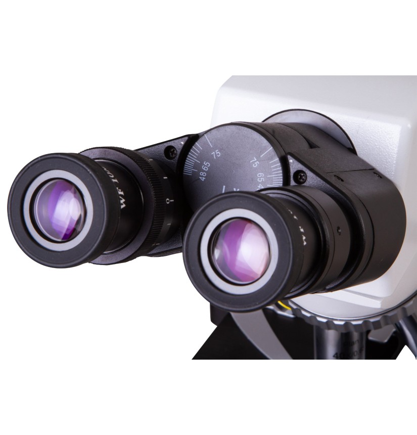
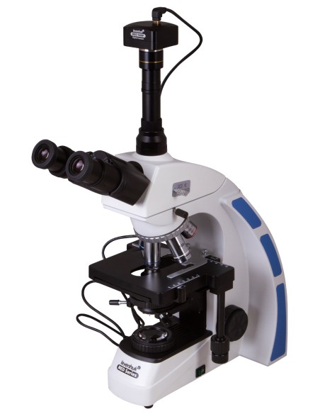
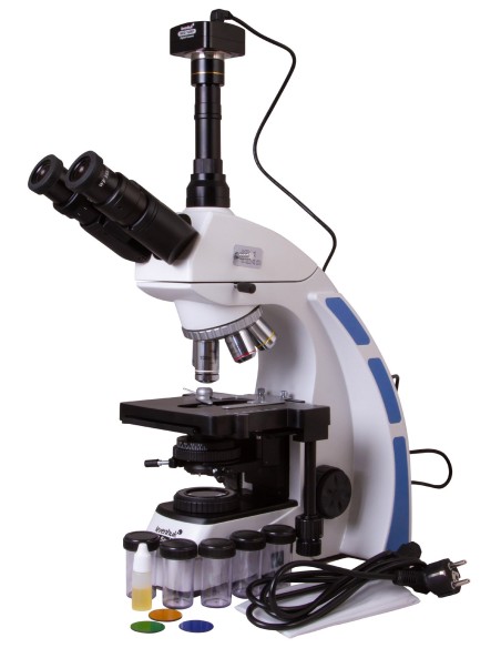
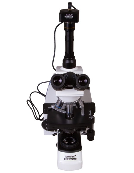
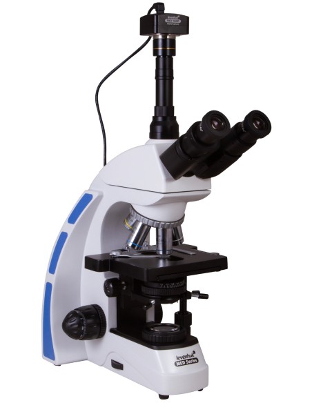
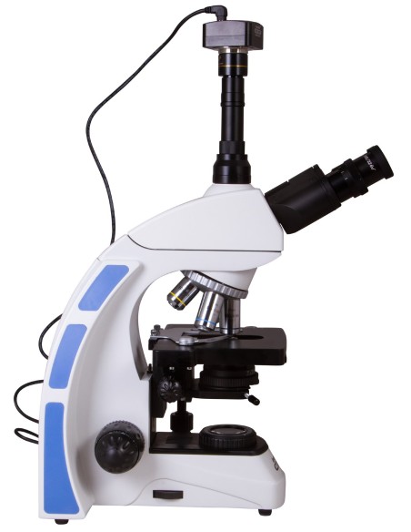
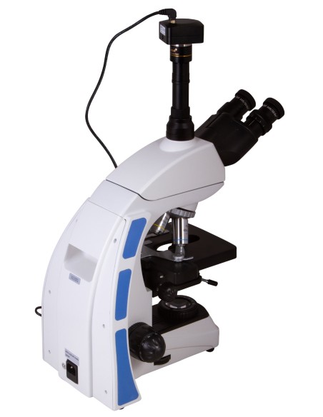
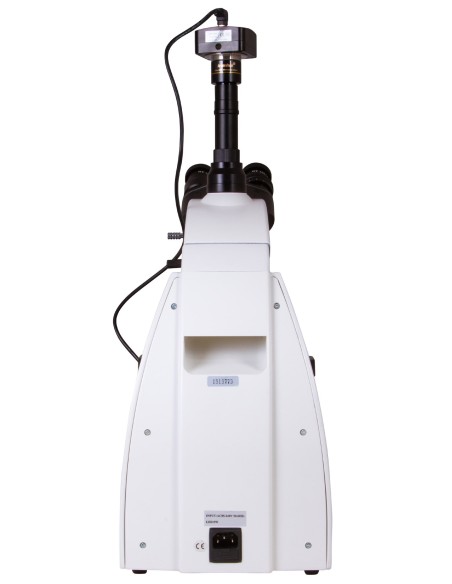
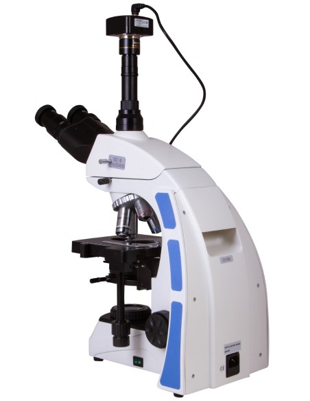
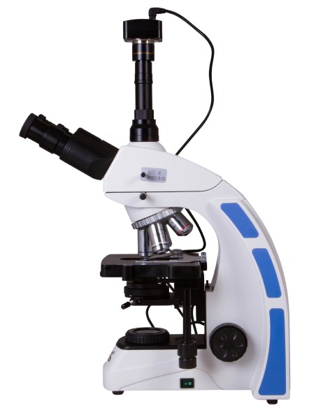
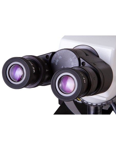


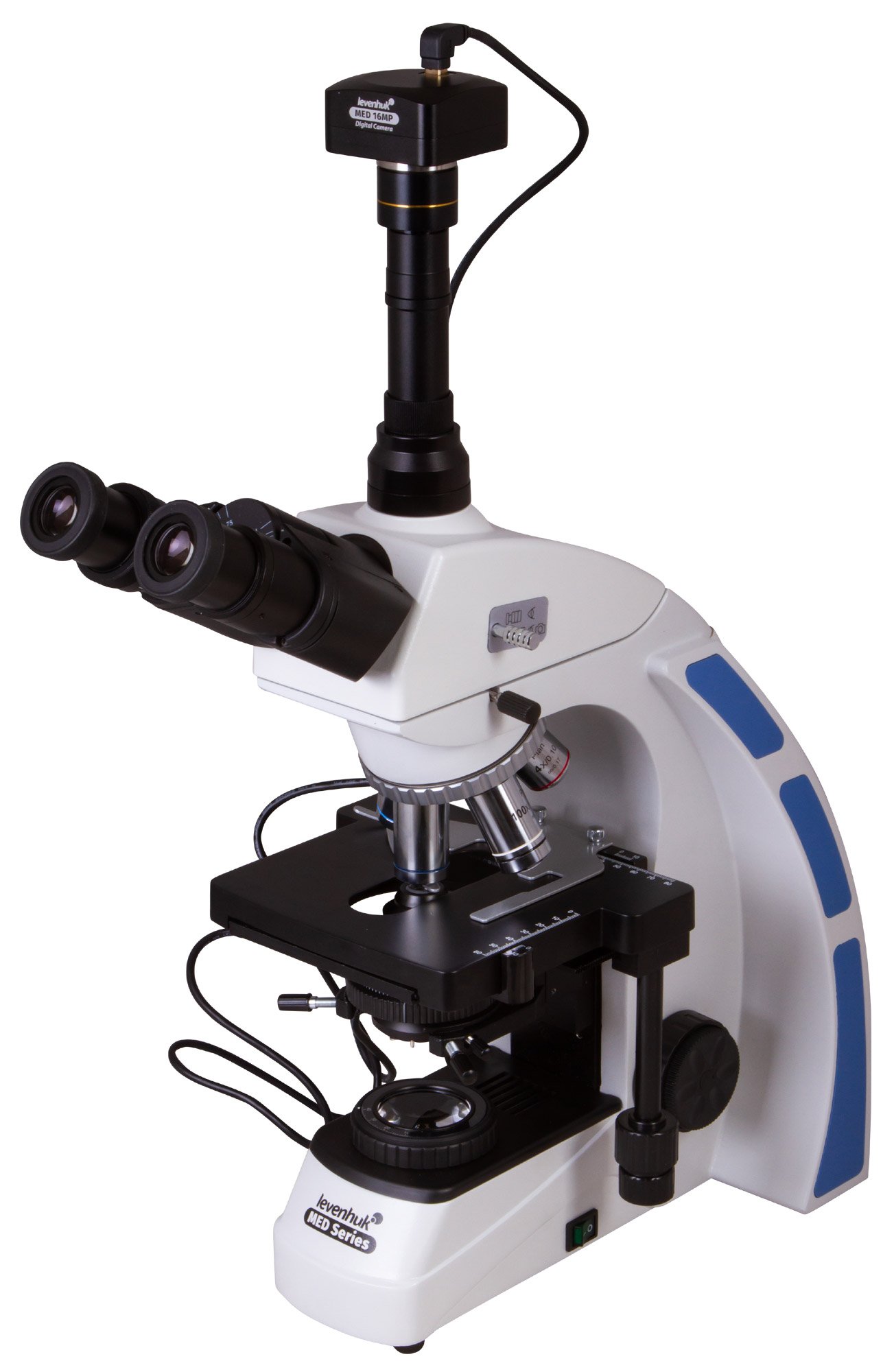
The Levenhuk MED D40T is a digital microscope with a trinocular head and a 16 MP camera included in the kit. The infinity-corrected planachromatic optical system produces a flat field, eliminates chromatic aberration and enables observations with a high level of detail. Köhler illumination can be used with the microscope, and oil immersion is available. The Levenhuk MED D40T is the perfect optical instrument for professionals who require a microscope with extensive functionality and high-quality optics.
Planachromatic optics with infinity correction
The Levenhuk MED 40 series microscopes are equipped with an infinity-corrected optical system, as used in professional and high-end microscopes. This system comprises planachromatic objectives and enables the transmission of clear, high-contrast images with excellent flatness.
One of the most important features of the infinity-corrected optical system is that it allows additional components to be fitted into the optical path between the objective lens and the eyepiece. These additional components include a polariser, epi-fluorescence unit and phase-contrast devices. Overall, the modular design and ease of use make Levenhuk MED 40 microscopes ideal instruments for use across various fields of microscopy, including work in haematology, histology, microbiology and many other laboratories.
Rotating head with beam splitter
The trinocular head is standard on microscopes supplied with a digital camera. The binocular section is used for visual observations, whilst the camera is housed in a separate eyepiece tube. There is no need to interrupt your work to take a photograph or record a video of samples. The visual module is slightly tilted at 30°, thereby preventing neck muscle stiffness during long hours of observation. The head can be rotated through 360°, and a beam splitter is included.
Optical capabilities
The kit includes optical accessories such as wide-field eyepieces with dioptre adjustment and planchromatic objective lenses of various magnifications. The microscope’s total magnification ranges from 40x to 1000x. Three high-magnification objectives have spring-loaded mounts. A 100x objective is designed for oil immersion. All optical components are protected by an anti-fungal coating.
16-megapixel digital camera for capturing photos and videos
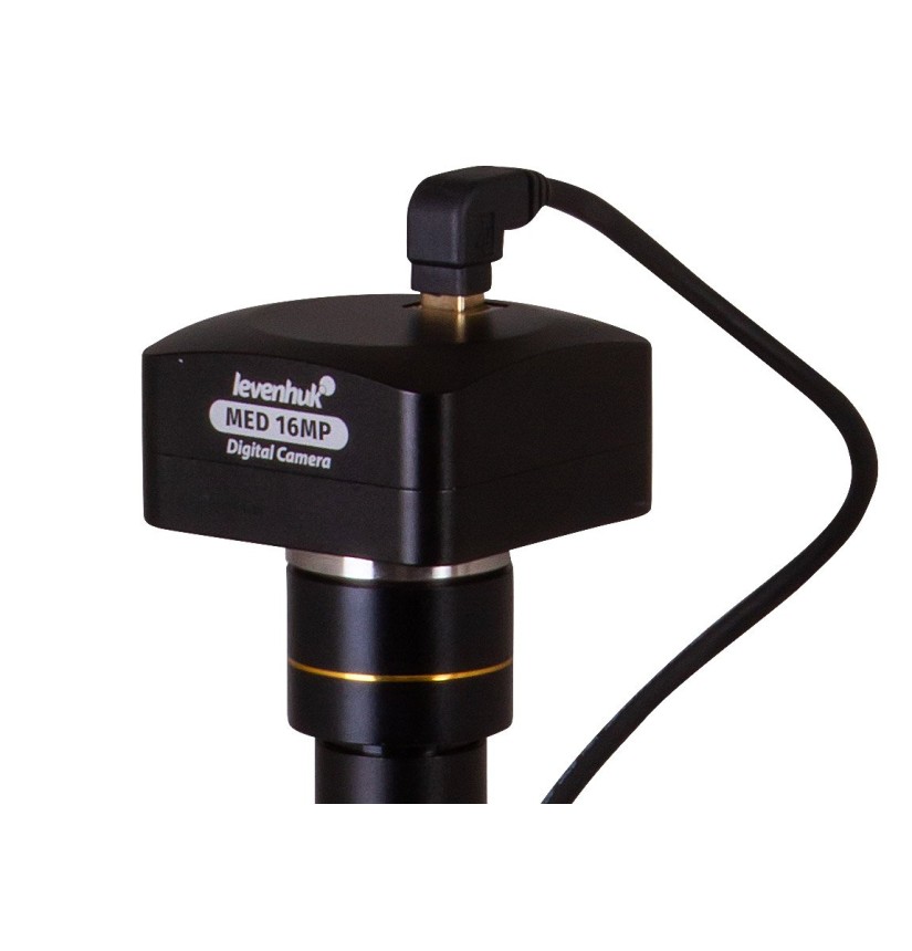
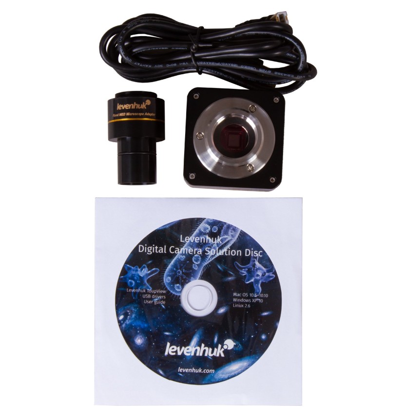
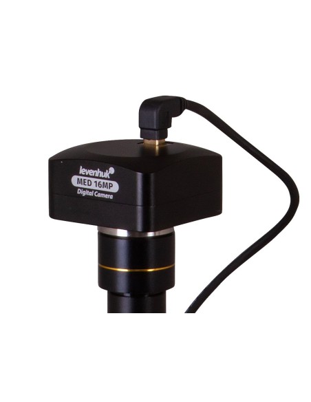
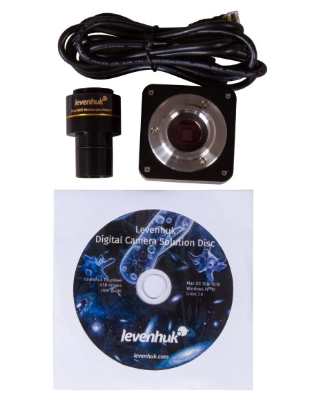
A 16 MP digital camera allows you to take high-resolution photos and record videos. The resulting images can be used for professional work with micro-samples and for creating digital archives of studies. The camera is connected to a PC via a USB cable (included in the kit) and transmits the image to the screen in real time. The camera transmits the image to the screen of a connected computer. This makes laboratory work simpler and more convenient, whilst also reducing strain on the eyes and shoulder joints, as there is no longer any need to look through eyepieces. Basic processing of the recorded content can be carried out using specialised software (the CD is included in the kit). The software allows you to adjust the image size, brightness and contrast, exposure time and white balance, calibrate the camera and lenses, as well as measure samples and structures (various units of measurement available).
Working comfortably with microscope slides
Samples are positioned under the objective lens using a mechanical stage. The stage can move in two directions. Coarse and fine focus are used to adjust the sharpness. A bright LED light is positioned on the base. The illumination system also includes an Abbe condenser with an iris diaphragm and a filter holder. Köhler illumination is available.
The light is powered by an AC power supply. The brightness of the light is adjustable.
Features:
- Planachromatic optics corrected to infinity
- Magnification range of the trinocular head with beam splitter from 40x to 1000x
- Intense LED light with adjustable brightness
- Köhler illumination available
- Powered by an AC adaptor, with adjustable brightness
- A 16 MP digital camera is included
The kit comprises:
- Microscope base with stand
- 360° rotatable trinocular head
- Infinity-corrected planachromatic objective lenses: 4X, 10x, 40xs, 60xs, 100xs (oil) with anti-fungal coating
- Wide-field eyepieces: WF10x/22 mm with anti-fungal coating (2 pcs.)
- Abbe condenser, N.A. 1.25, with iris diaphragm and filter holder
- Filters: blue, green, yellow
- Bottle of immersion oil
- Fuses (2 pcs.)
- Microscope power cable
- Dust cover
- 16 MP digital camera
- Camera adapter
- Camera mount
- USB cable for connecting and charging the camera
- CD with software and drivers
- User manual and lifetime warranty
Warning:
for information on the correct mains voltage, please refer to the specifications table. Never attempt to connect a 110 V device to a 220 V mains socket, or vice versa, without using a voltage converter. Please note that the mains voltage is 220–240 V in most European countries and 110 V in the United States and Canada.
Some things you can observe under the microscope:


.
The Levenhuk MED D40T digital trinocular microscope is compatible with Levenhuk digital cameras (sold separately). Levenhuk cameras are fitted onto the optical tube in place of an eyepiece.
| Brand | Levenhuk Inc., USA |
| Warranty, years | Lifetime |
| Package dimensions (WxDxH), cm | 50x44x28 |
| Shipping weight, | 10.34 |
| Type | organic |
| Eyepiece | binocular |
| Optical material | optical glass with anti-fungal coating |
| Nozzle | rotates 360° |
| Head tilt angle | 30° |
| Magnification, x | 40–1000 |
| Eyepiece tube diameter, mm | 30 mm (binocular head), 23.2 mm (third vertical tube) |
| Eyepieces | WF10x/22 mm, wide-field with dioptre adjustment (2 pcs.) |
| Objectives | planachromatic objectives corrected to infinity: 4x, 10x, 40xs, 60xs, 100xs (oil) |
| Objective turret | for 5 objectives |
| Interpupillary distance, mm | 48–75 |
| Stage, mm | 180 × 150 |
| Plate movement range, mm | 75 × 50 |
| Plate features | double mechanical layer, with mechanical scale |
| Dioptre adjustment of the eyepiece, dioptres | ±5 |
| Condenser | Abbe N.A. 1.25 with iris diaphragm and filter holder |
| Iris diaphragm | iris |
| Focus | coaxial, macrometric (0.5 mm) and micrometric (0.002 mm), with rack and pinion |
| Body | metal |
| Illumination | LED |
| Brightness adjustment | True |
| Power supply | 100–240V |
| Light source type | 5W, 85–230 V AC |
| Optical filters | green, blue, yellow |
| Megapixels | 16.0 |
| Sensor element | 1/2.33” (6.18 × 4.66 mm) |
| Pixel size, µm | 1,335 × 1,335 |
| Video recording | Yes |
| Frame rate | 2@4632x3488 8@2320x1740 11@1536x1160 |
| Position for use | the microscope’s third 23.2 mm eyepiece tube; instead of the eyepiece |
| Image format | *.jpeg |
| Spectral range, nm | 380–650 |
| White balance | automatic/manual |
| Exposure control | 0.2–2000 μs |
| Software, drivers | Levenhuk |
| Programmable options | image size, brightness, exposure time |
| Output | USB 2.0, 480 Mbps |
| System requirements | Windows 7/8/10 (32-bit and 64-bit), compatible with Mac OS 10.6–10.10 and Linux, processor up to 2.8 GHz Intel Core 2 or higher, 2 GB RAM or more, USB port, CD-ROM |
| Camera power supply | via USB cable |
| Application | laboratory/medical |
| Lighting position | bottom |
| Search method | brightfield |
| Digital camera included | True |