








































































Your cart
There are no more items in your cart


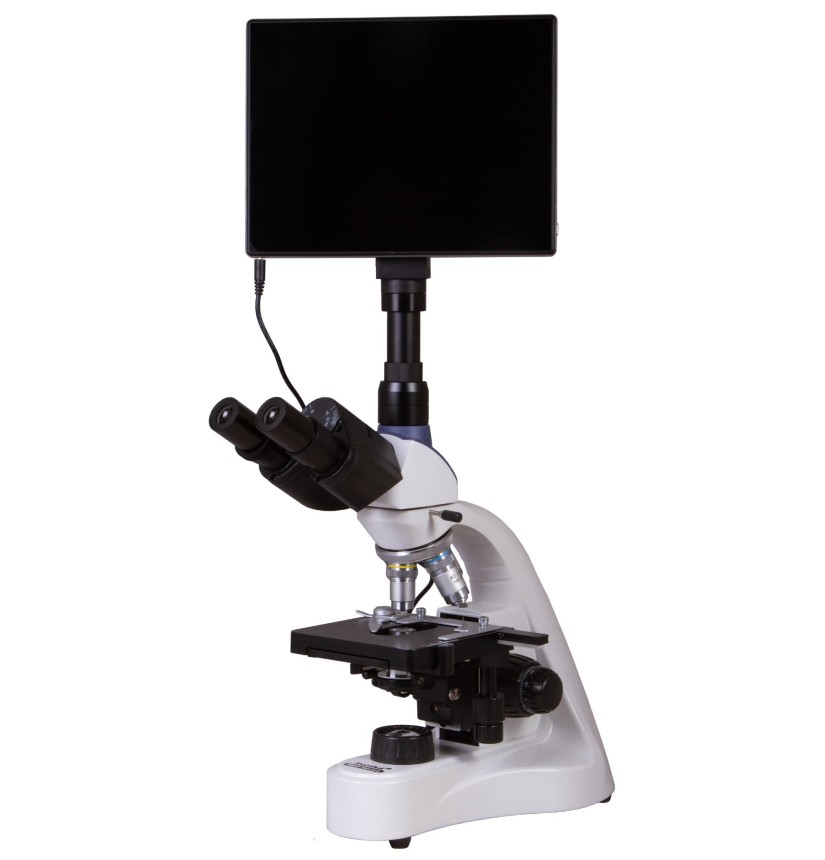
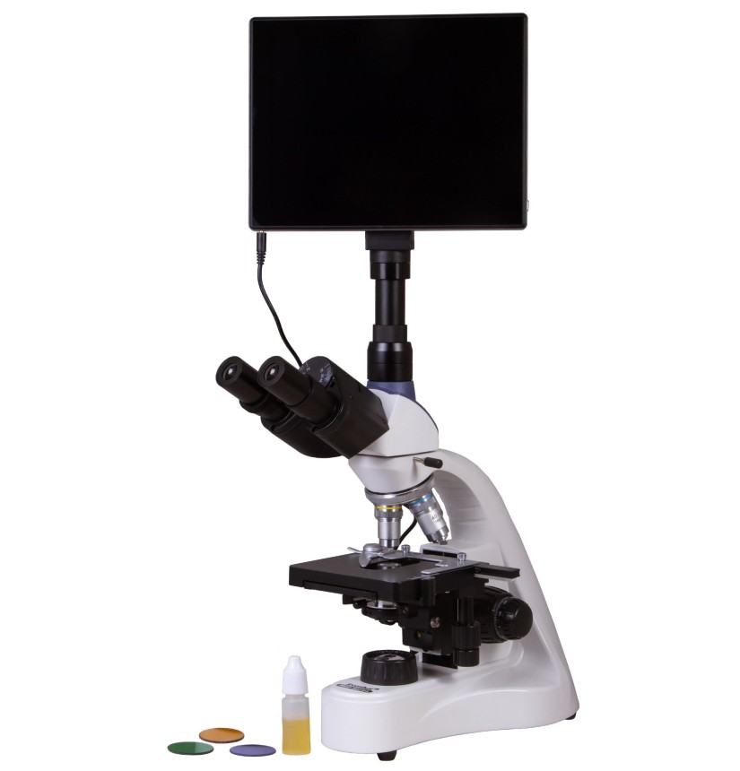
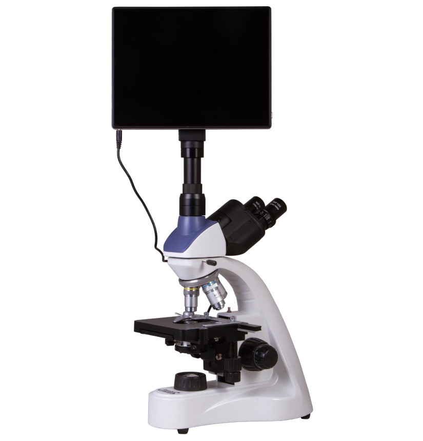
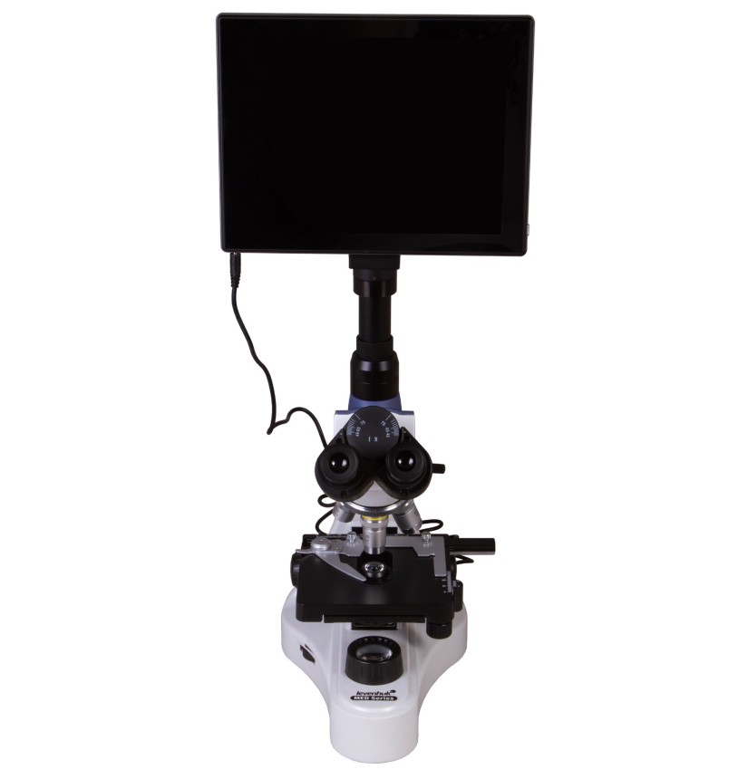
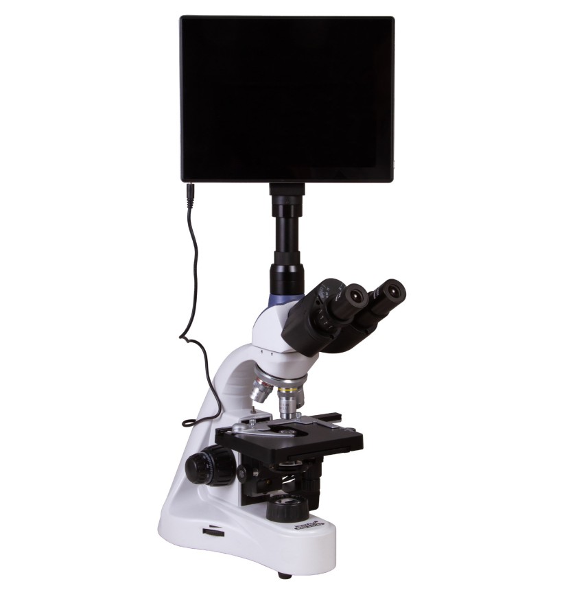
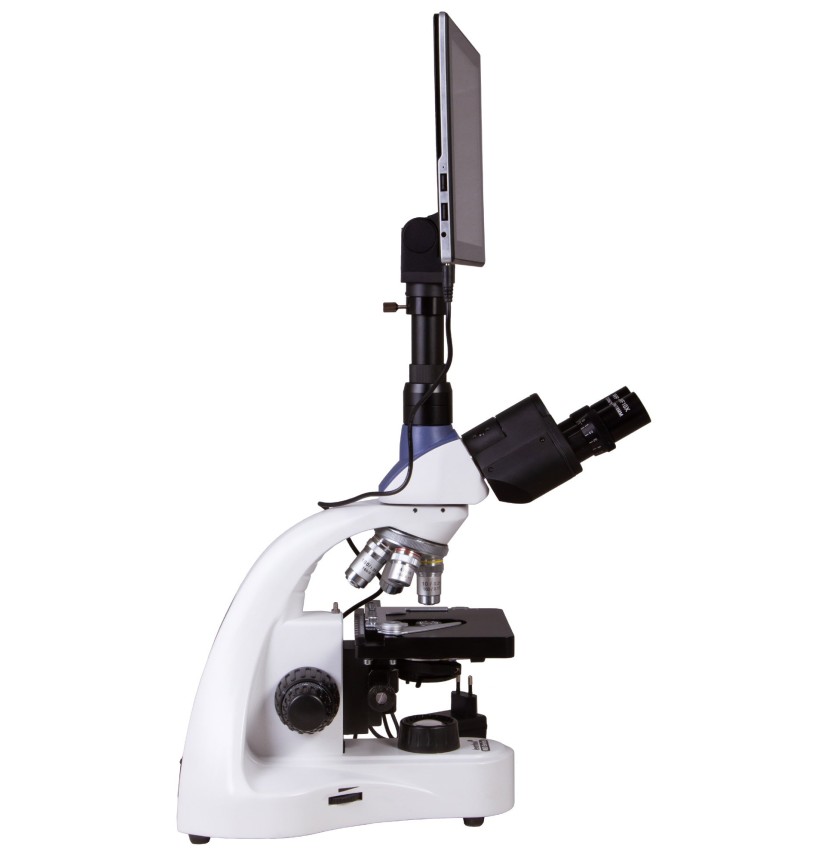
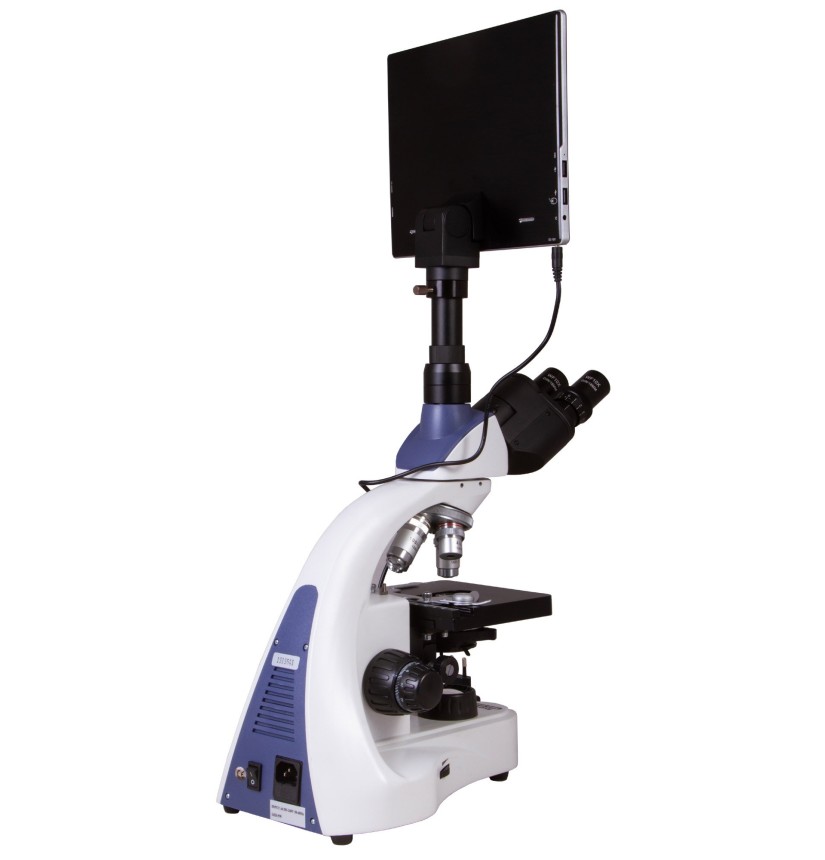
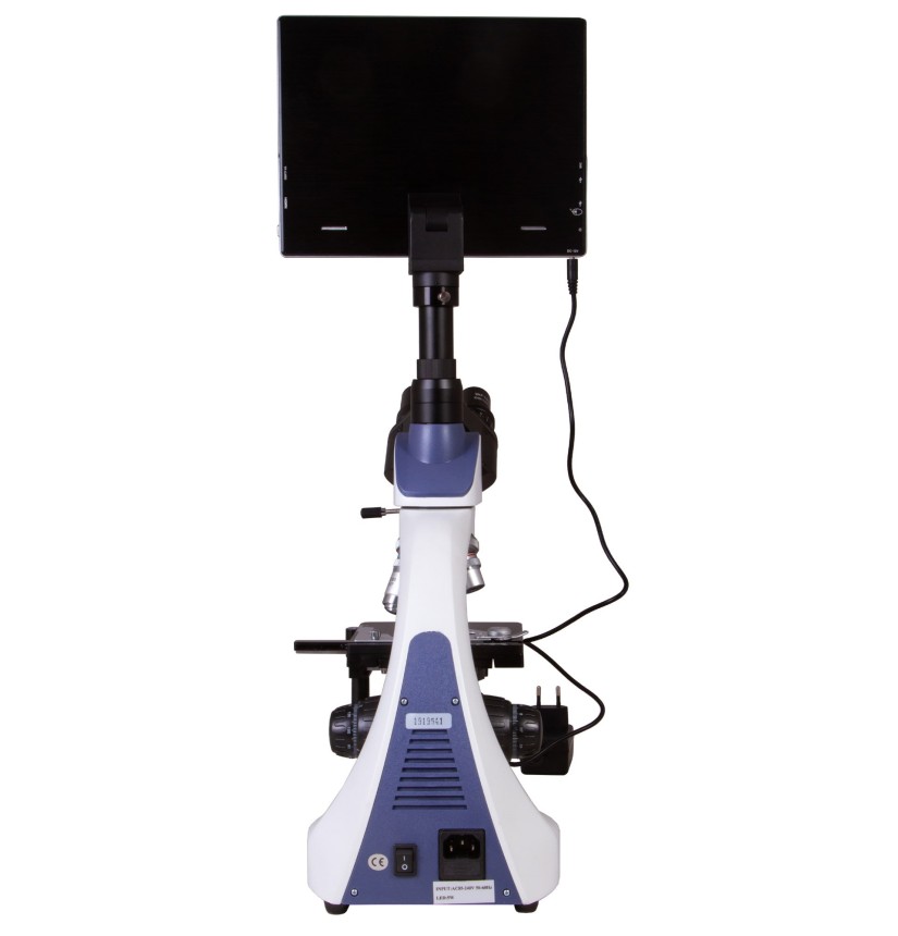
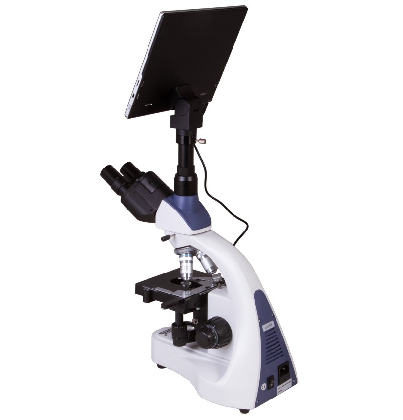
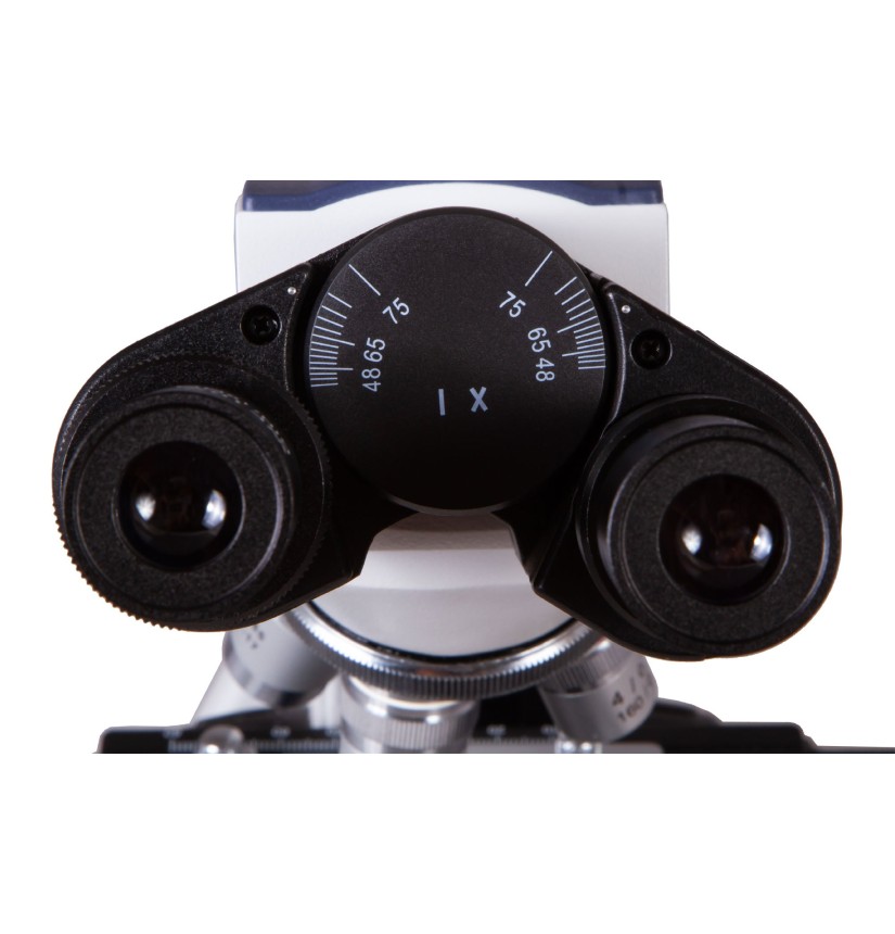
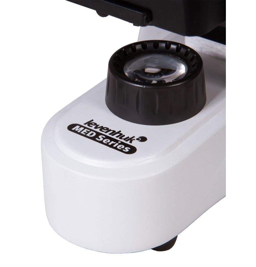
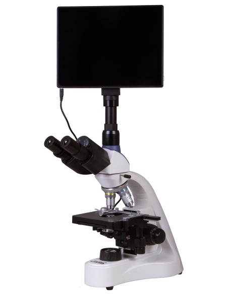
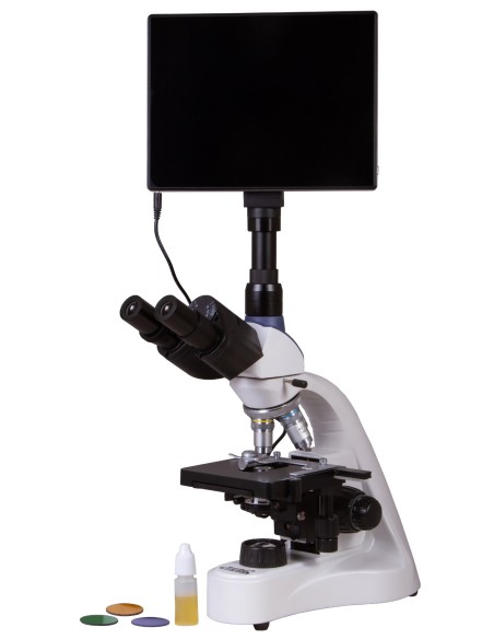
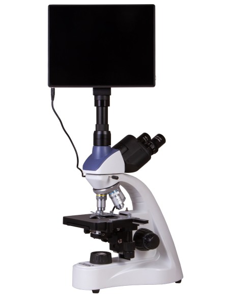
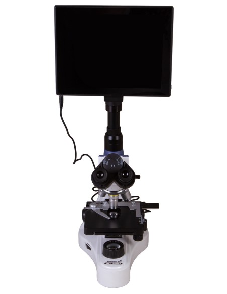
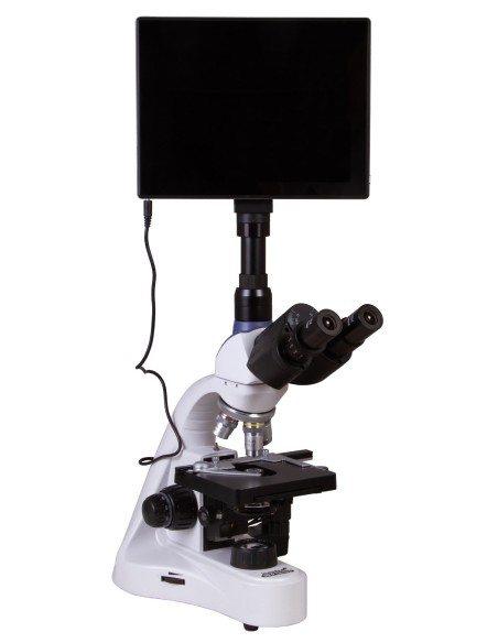
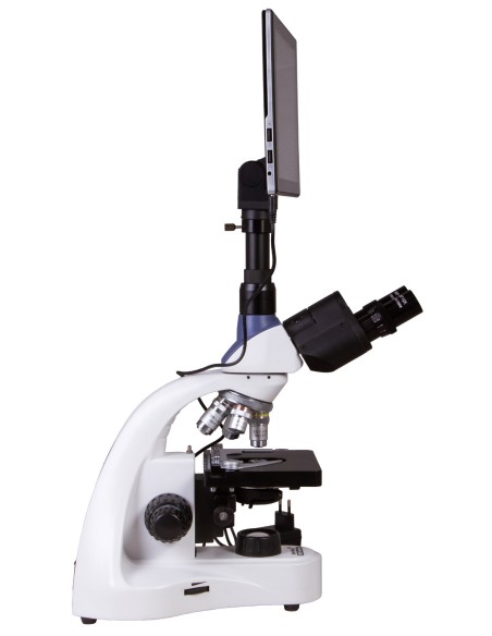
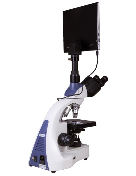
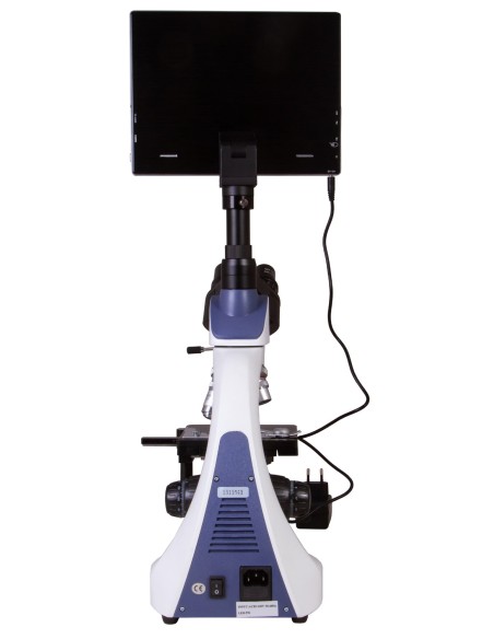
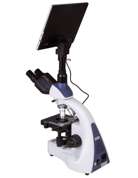
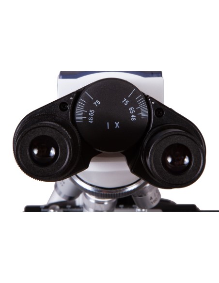
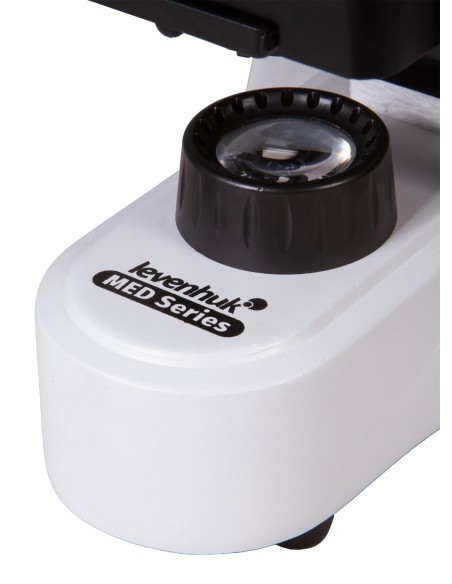


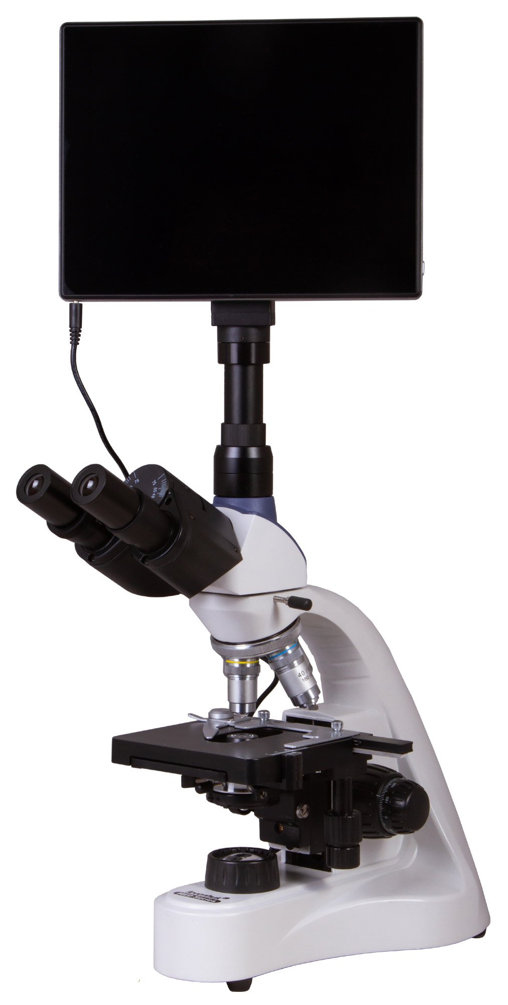
The Levenhuk MED D10T LCD digital microscope is equipped with a digital camera and LCD screen, meaning it does not need to be connected to a computer. As a result, the microscope is self-contained: no additional equipment is required to make full use of its capabilities. It allows you to carry out observations using the bright-field method, take research photographs and record them as video. The image resolution is sufficient to provide a sharp and clear view of the fine details of complex biological structures.
High-quality optics
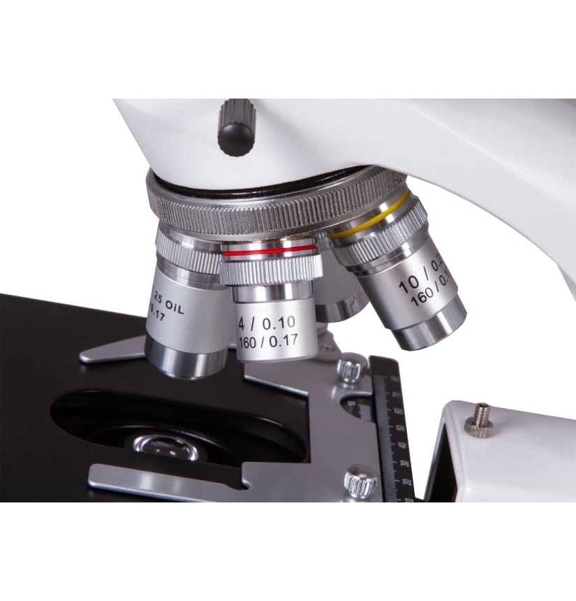
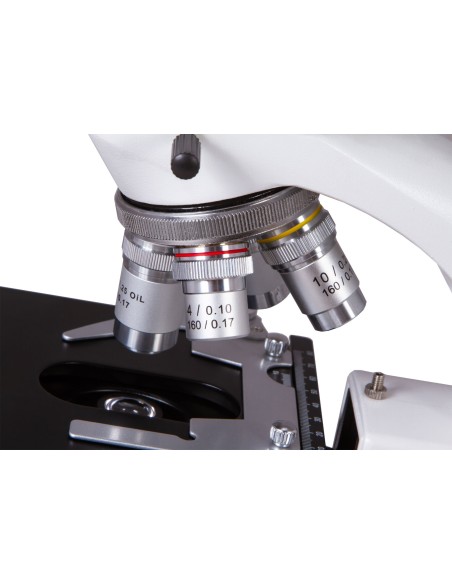
The achromatic objective lenses allow for detailed observation of micro-objects with a magnification range from 40x to 1000x. The 100x objective lens can be used for observations using oil immersion (a bottle of immersion oil is included in the kit). Furthermore, high-magnification objectives feature spring-loaded mounts that protect against accidental damage. The wide-field eyepieces provide 10x magnification and are suitable for working with large specimens. All optical elements are protected by an anti-fungal coating.
Practical swivel head
The head consists of a binocular viewing unit and a vertical eyepiece tube, in which a digital camera is installed. A 30° tilt angle on the viewing unit is comfortable for prolonged observations and helps reduce strain on the neck muscles. The 360° rotatable head is useful for group research.
Digital capabilities
The digital camera deserves special mention: a 5 MP sensor allows you to take high-quality photos and record videos; the image seen through the lens is transmitted to an LCD screen in real time. The pre-installed software allows you to configure the camera, edit photographs and videos, and save images for further processing. You can also connect headphones, a keyboard and a mouse. External storage is supported. A camera adapter for attaching the camera to the eyepiece is included in the pack. The camera makes laboratory work easier and more convenient for researchers: there is no need to strain your eyes to view slides through the eyepieces, as everything is visible on the integrated LCD screen with touch controls. The software allows you to adjust the image size, brightness and contrast, gamma, sharpness and saturation, exposure time and white balance by calibrating the camera and objective lenses; it also enables you to measure samples and structures (various units of measurement available). In addition, the programme enables you to carry out particle size analysis.
Working comfortably with microscope slides
The stage is fitted with a mechanical scale, which is used for the precise positioning of samples under an objective lens. A double-layer mechanical stage can be moved along two axes. Coarse and fine focus are used to adjust the sharpness.
Intense illumination of slides
The illumination system is located at the base of the microscope. It consists of a powerful 5 W LED light and an Abbe condenser with an iris diaphragm and filter holder. The brightness of the light is adjustable. It is powered by an AC power supply.
Features:
• Laboratory microscope with
40–1000x magnification• 360° rotatable trinocular head, inclined at
30°• Achromatic optics with anti-fungal coating•
5 W lower LED light with brightness control•
Powered by an AC power supply•
The kit includes a 5 MP digital camera and an LCD screen with Android
operating system
The kit includes:
• Microscope base with
stand• 360° rotatable trinocular head•
Achromatic objective lenses: 4×, 10×, 40×s, 100×s (oil) with
anti-fungal coating• WF10×/18 mm wide-field eyepieces with anti-fungal coating (2 pcs.)
• Abbe condenser N.A. 1.25 with iris diaphragm and filter holder•
Filters: blue, green,
yellow• Bottle of
immersion oil• Fuse (2 pcs.)
• Microscope power cable•
Dust cover•
5 MP digital camera with LCD screen•
Camera adapter• Camera power cable•
User manual and lifetime
warranty
Warning:
For information on the correct mains voltage, please refer to the specifications table; never attempt to connect a 110 V device to a 220 V mains socket, or vice versa, without using a converter. Please note that the mains voltage is 220–240 V in most European countries and 110 V in the United States and Canada.
Some things you can observe with a microscope:


The Levenhuk MED D10T LCD digital trinocular microscope is compatible with Levenhuk digital cameras (sold separately). Levenhuk cameras should be fitted onto the eyepiece tube, in place of an eyepiece.
| Brand | Levenhuk Inc., USA |
| Warranty, years | Lifetime |
| Package dimensions (LxWxH), cm | 57x38x26 |
| Shipping weight, | 6.66 |
| Head | trinocular |
| Optical material | optical glass with anti-fungal coating |
| Nozzle | rotates 360° |
| Head tilt angle | 30° |
| Magnification, x | 40–1000 |
| Eyepiece tube diameter, mm | 30 mm (binocular head), 23.2 mm (third vertical tube) |
| Eyepieces | 10x/18 mm, wide-field with dioptre adjustment (2 pcs.) |
| Objectives | achromatic: 4x, 10x, 40xs, 100xs (oil immersion) |
| Interpupillary distance, mm | 48–75 |
| Stage, mm | 125 × 130 |
| Stage features | double mechanical layer, with mechanical scale |
| Eyepiece dioptre adjustment, dioptres | ±5 |
| Condenser | Abbe N.A. 1.25 with iris diaphragm and filter holder |
| Iris diaphragm | iris |
| Focus | coaxial; coarse (20 mm) and fine (0.02 mm) |
| Body | metal |
| Illumination | LED |
| Brightness adjustment | True |
| Power supply | 100–240V |
| Light source type | 5 W |
| Optical filters | green, blue, yellow |
| Megapixels | 5.0 |
| Sensor element | 1/2.5” |
| Pixel size, µm | 2.2 × 2.2 |
| Sensitivity, V/lux-sec@550 nm | 0.53 |
| Video recording | yes |
| Frame rate | 15 frames per second |
| White balance | automatic/manual |
| Exposure control | automatic/manual |
| Software, drivers | Android 5.1 (multilingual) |
| Programmable options | measurement, brightness, particle size analysis, etc. |
| Output | USB 2.0 (2 PCs), mini HDMI, Wi-Fi, TF memory card slot |
| Camera power supply | 12 V AC, via AC adaptor |
| Special features | 9.4-inch colour LCD screen with sensor; built-in memory: 4 GB |
| Option to connect additional equipment | Support for microSD cards with capacities up to 32 GB; monitor/TV (via HDMI port); flash drive, computer mouse, keyboard (via a USB connector); headphones (3.5 mm) |
| Image | *.jpeg |
| Video format | 1080p, *.3gp |
| Application | laboratory/medical |
| Search method | brightfield |
| Digital camera included | True |
| Maximum resolution | 2048x1536 |